Image Credit: Shutterstock Image
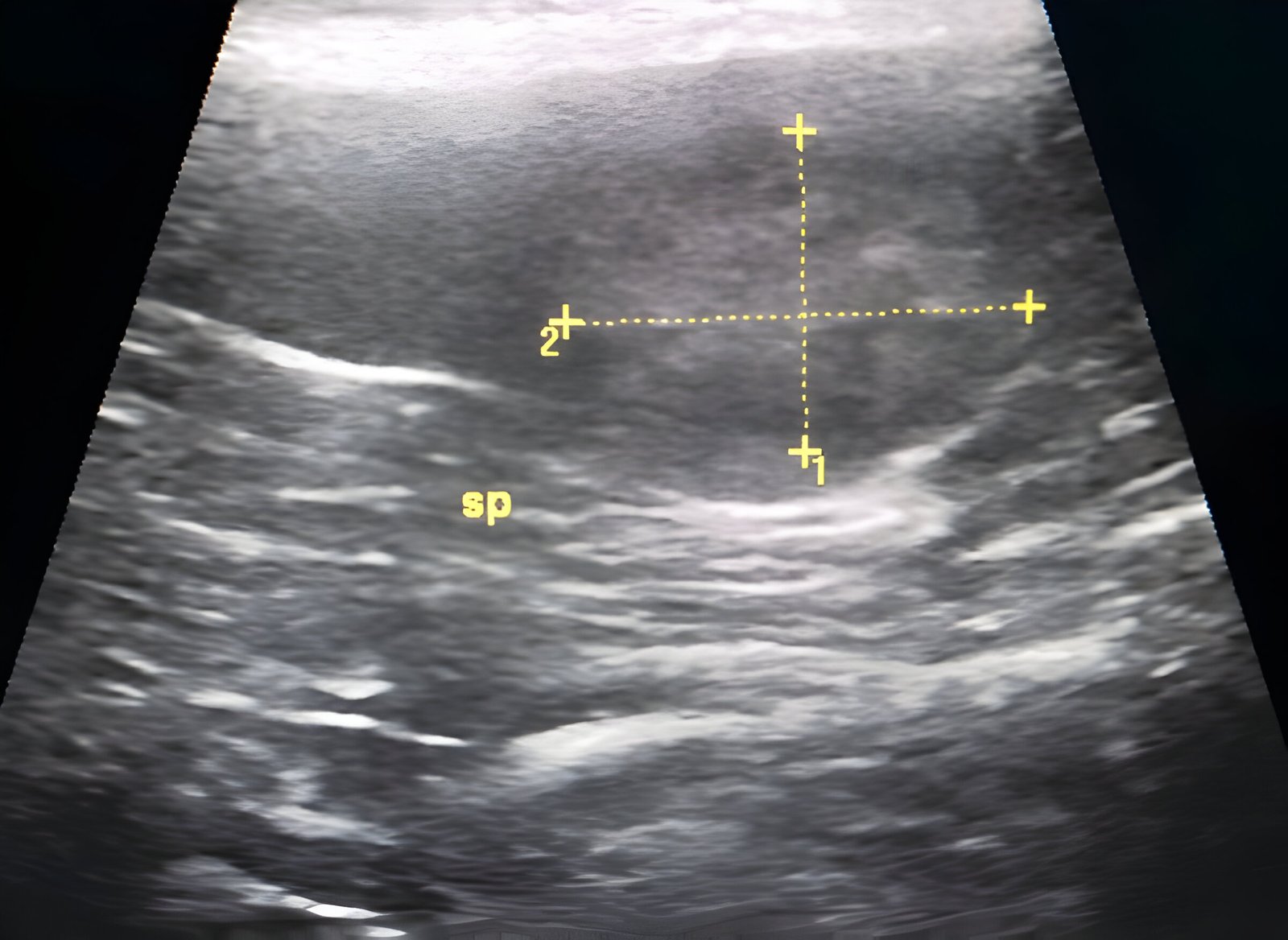
Medical imaging plays a crucial role in diagnosing various health conditions, and one common finding that often raises concerns is a hypoechoic mass. This term refers to a structure or lesion that appears darker than the surrounding tissue on an ultrasound image. Hypoechoic masses can occur in different parts of the body, including the breast, thyroid, and kidney, and their detection has an impact on patient care and treatment decisions.
Understanding the characteristics and implications of hypoechoic masses is essential for healthcare providers and patients alike. This article aims to explore the causes, symptoms, and treatment options associated with hypoechoic masses. It will delve into the specifics of breast ultrasound imaging, discuss how to tell apart benign and malignant masses, and outline the approaches to manage these findings. By shedding light on this topic, we hope to provide valuable insights to help readers better understand this common medical phenomenon.
Read More: What Causes Back Pain In Females
Table of Contents
ToggleUnderstanding Breast Ultrasound Imaging
How ultrasound works
Breast ultrasound imaging uses high-frequency sound waves to produce pictures of the internal structures of the breast. The process involves a handheld device called a transducer, which emits sound waves that bounce off breast tissues. These echoes are then captured and transformed into images on a computer monitor. This technology operates on the same principles as sonar used by bats and ships, allowing healthcare providers to visualize the size, shape, and consistency of breast tissues.
Interpreting ultrasound images
Interpreting breast ultrasound images requires expertise and experience. Radiologists analyze these images to detect changes in the appearance of organs, tissues, and vessels, as well as to identify abnormal masses such as tumors. Ultrasound has an impact on differentiating between fluid-filled cysts and solid masses, which is crucial for determining whether a lump is potentially cancerous. Additionally, Doppler ultrasound techniques can be employed to evaluate blood flow within breast masses, providing valuable information about their nature.
Advantages and limitations
Breast ultrasound offers several advantages. It is safe, painless, and does not use ionizing radiation, making it suitable for pregnant women and those who cannot undergo mammography. Ultrasound is particularly useful for examining dense breast tissue, where mammograms may have limitations. It also serves as a valuable tool for guiding needle biopsies and assessing tumor response to chemotherapy.
However, breast ultrasound has limitations. It may miss some early signs of cancer, such as microcalcifications, which are better detected by mammography. The accuracy of ultrasound can be affected by factors such as the patient’s body composition and the operator’s skill level. Therefore, it is crucial to choose a facility with expertise in breast ultrasound, preferably one where radiologists specialize in breast imaging.
Characteristics of Hypoechoic Masses
Shape and margins
Hypoechoic masses can have various shapes and margins, which play a crucial role in determining their nature. The shape of a mass can be oval, round, or irregular. Oval masses are elliptical or egg-shaped, sometimes including two or three undulations. Round masses appear spherical, ball-shaped, or circular. Irregular masses have neither a round nor oval shape and often raise suspicion for malignancy.
The margins of hypoechoic masses can be circumscribed, obscured, microlobulated, indistinct, or spiculated. Obscured margins are partially hidden by adjacent fibroglandular tissue. Microlobulated margins show short-cycle undulations, while indistinct margins lack clear demarcation.
Echo patterns
Hypoechoic masses appear darker or more gray than the surrounding tissue on ultrasound images. This is because they absorb more ultrasound waves and are typically composed of dense tissue, such as muscle or fibrous connective tissue. In contrast, hyperechoic masses appear lighter or brighter, often indicating air-, fat-, or fluid-filled structures.
Size and orientation
The size and orientation of hypoechoic masses are important factors in their assessment. Masses that are 2 centimeters or larger and contain calcium deposits are more likely to be cancerous. However, orientation alone should not be the sole determining factor when assessing malignancy risk.
Click For More: Psoas Stretch
Differentiating Benign vs. Malignant Masses
Key ultrasound features
Ultrasound imaging plays a crucial role in distinguishing between benign and malignant masses. Benign tumors typically have regular contours, acoustic shadows, and low Doppler color in septae. In contrast, malignant masses often exhibit irregular contours, absence of acoustic shadows, and the presence of solid areas with moderate to high Doppler color. The shape, margins, and echo patterns of hypoechoic. masses provide valuable information.
BI-RADS classification
The Breast Imaging Reporting and Data System (BI-RADS) offers a standardized approach to categorize mammogram findings. BI-RADS 2 indicates benign findings with a 0% probability of malignancy.
Additional diagnostic tools
To enhance diagnostic accuracy, healthcare providers employ additional tools. Contrast-enhanced ultrasound (CEUS) assesses blood flow in lesions through perfusion imaging of small capillaries. Ultrasound elastography evaluates tissue stiffness, offering improved specificity and accuracy in differentiating benign from malignant breast tumors. However, elastography results can be influenced by operator skills and lesion location. In some cases, sono-elastography has the potential to downgrade BI-RADS category 4a lesions to category 3, potentially reducing unnecessary biopsies.
Management of Hypoechoic Masses
The management of hypoechoic masses depends on various factors, including the type, size, location, and symptoms associated with the mass. Healthcare providers employ different approaches based on the specific characteristics of the mass and the patient’s overall health.
Watchful waiting
In some cases, doctors may opt for a watchful waiting approach. This strategy involves carefully monitoring the mass without immediate intervention. It is often employed when:
- The mass appears benign and does not cause significant symptoms.
- The underlying condition causing the mass may resolve on its own.
- The risks of intervention outweigh the potential benefits.
During this period, healthcare providers may recommend follow-up ultrasound scans to track any changes in the mass’s size or appearance.
Minimally invasive procedures
For certain hypoechoic masses, minimally invasive procedures offer effective treatment options. These techniques aim to shrink or remove the mass with minimal disruption to surrounding tissues. Some common minimally invasive procedures include:
- Radiofrequency ablation: This technique uses electrical currents to shrink masses.
- Biopsy: While primarily diagnostic, some biopsies can also serve as a treatment method for smaller masses.
Surgical options
Surgery often serves as the primary treatment for larger hypoechoic masses or those suspected of malignancy. Surgical intervention has an impact on:
- Removing benign growths that cause pain, obstruction, or other complications.
- Excising masses that affect organs, blood vessels, or nerves.
- Addressing cosmetic concerns related to visible masses.
Surgical approaches vary depending on the mass’s location and characteristics. They may include:
- Keyhole or laparoscopic procedures: These involve small incisions and are less invasive.
- Endoscopic techniques: These allow access to internal organs without external incisions.
- Traditional open surgery: This may be necessary for larger or more complex masses.
Conclusion
Hypoechoic mass are a common finding in medical imaging that can have a big impact on patient care. These darker areas on ultrasound scans can show up in different parts of the body and may point to various health issues. By looking at things like shape, size, and echo patterns, doctors can often tell if a mass is likely to be harmless or possibly dangerous. This helps them decide whether to keep an eye on it, do more tests, or start treatment right away.
In the end, dealing with hypoechoic masses depends on each patient’s situation. Sometimes, doctors choose to watch and wait, especially if the mass seems harmless. Other times, they might use minimally invasive procedures or surgery to get rid of the mass. The key is to work closely with healthcare providers to figure out the best approach for each case, balancing the need to address potential health risks with the goal of avoiding unnecessary procedures.
FAQs
What should I do if I find a hypoechoic heterogeneous mass? If you discover a hypoechoic heterogeneous mass, especially during a breast sonogram, it’s important to take it seriously as it can sometimes indicate cancer, though it could also be benign. Sonogram technicians employ specific techniques to differentiate between benign and malignant masses, and in some cases, a biopsy may be necessary to determine the nature of the mass.
What are the possible causes of a hypoechoic breast mass? Hypoechoic breast masses can arise from a variety of causes. Benign conditions such as fibroadenomas, cysts, and intramammary lymph nodes frequently cause these masses. However, they can also indicate the presence of breast cancer, particularly invasive ductal carcinoma.
Why are some structures described as hypoechoic on ultrasound scans? A structure is termed hypoechoic when it reflects fewer ultrasound waves compared to the surrounding tissues. The prefix ‘hypo-‘ means less than normal, and ‘-echoic’ relates to echoes, or the sound waves used in ultrasound imaging. On an ultrasound image, these hypoechoic structures appear darker than other areas.
What are the treatment options for a hypoechoic mass? Treatment for a hypoechoic mass may often involve surgery, particularly if the mass is large. Even benign masses can lead to complications such as pain, obstruction, or even the risk of becoming cancerous or causing internal bleeding due to rupture. Surgical removal is commonly considered when masses impact organs, blood vessels, or nerves.


Leave a Reply